Key learning objective
To review the general design features of the slit lamp
History
Over a century of biomicroscopy:
1891 Aubert first used a binocular microscope
1897 Czapski & Zeiss developed the instrument further
Principle
A narrow "slit" beam of very bright light produced by lamp. This beam is focused on to the eye which is then viewed under magnification with a microscope
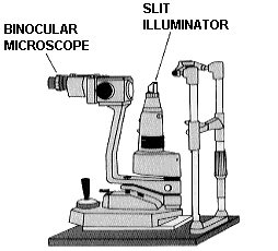
Components of the slit lamp
The illumination system
The microscope
The remainder of the instrument (e.g., chin rest, coupling etc.)
from Henson (1983)
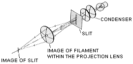
The illumination system
The aim of the designers is to produce a uniformly bright, accurately focused slit of light whose dimensions can be adjusted. This is usually obtained by using the Vogt-Koeller projection system.
The distance between projector lens and focused beam (i.e., the focal length of the projector lens) is short to maximise illumination but not so short that there is no room for the practitioner to carry out manipulative procedures.
Room is required for attachments such as the applanation tonometer.
The illumination consists of:
- a light source
(usually halogen) often with a reflector positioned behind it to maximise illumination.
- condensing lenses
. Usually a pair of aspheric plano-convex lenses.
The light source is positioned at principal focus of the first lens. These lenses are aspheric to reduce chromatic aberration.
- a slit aperture or diaphragm to alter length, width and orientation of slit
- projector lens.
This is small to reduce aberrations.
The magnification/viewing system
Clinicians require a magnification from about x 10 to about x 50. A compound microscope is employed.
Methods of changing magnification include
- change objectives (e.g., Haag Streit)
- Littmann-Galilean telescope
Littmann-Galilean system (from Henson, 1983)
- Zoom systems Þ
gradual change in magnification
Inverting prisms are built in to "re-invert" the inverted image produced by the compound microscope.
Eyepieces/prisms can be rotated to change pd
A yellow barrier filter may be employed in the viewing system. Blue light from the cobalt blue filter (peak approx. 400 nm) in the illumination system is absorbed by fluorescein sodium (peak absorption approx. 480 nm) which then emits green light (peak approx. 550 nm). When the clinician views the eye there is rather poor contrast between the green fluorescence and the blue background light. A yellow barrier filter (absorbing wavelengths below approx. 510 nm) may be placed in the viewing system to remove all blue light. The green of the fluorescein then can be seen more easily.
General design features
The illumination and observation systems are coupled around a common centre of rotation to ensure that the microscope is always focused at the same point as where the slit beam is focused. A mechanism to enable the instrument to decouple (for e.g., sclerotic scatter) is required.
A joystick control is employed to enable instrument to be moved left-right/forward-backward (= focus)/ and up-down.
A chin rest, head rest and fixation target is also required. Some slit lamps have a tilting mechanism to enable the lamp to be directed from different angles.
Locking mechanisms are included.
Further reading
Henson DB (1983) Optometric Instrumentation. Chapter 6. Butterworths, Oxford